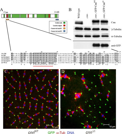
Figure 1.
Motif 1 of Cnn is essential. (A) Schematic of Cnn protein showing the two conserved motifs at the amino and carboxyl termini. An expanded view of Motif 1 is shown with the 13 amino acids that are deleted in CnnΔ1 underlined in red. The alignments include the putative Cnn homologues in Aedes egypti (mosquito, A.e.), CDK5RAP2 from Gallus gallus (chicken, G.g.) and human (H.s.), Myomegalin/PDE4DIP from human, and Mto1p from S. pombe (S.p.). (B) Western blot showing the expression of GFP-CnnWT and GFP-CnnΔ1 in a cnn null mutant (cnnhk21/cnn25cn1) background (labeled “cnn-” above the lanes). The samples were loaded onto two gels and blotted, and one gel was probed with anti-Cnn, anti-γ-Tub, and anti-α-Tub (loading control) antibodies as indicated. The second blot, shown below the solid line, was probed with anti-GFP antibody. Immunostaining of cnn null embryos expressing GFP-CnnWT (C) and GFP-CnnΔ1 (D) shows localization to centrosomes and mutant phenotype. Anti-GFP signal is green, anti-α-Tub signal is red, and DNA is blue. The erratic spacing of mitotic figures, linked spindles, and aneuploid nuclei in D are all characteristic of cnn mutant embryos, showing that GFP-CnnΔ1 did not rescue. Note the abundance of centrosome pairs or clusters in the cnnΔ1 mutant embryo.

