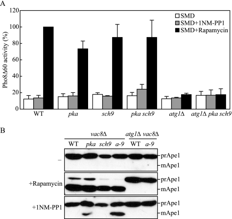
Figure 2.
Nonspecific and specific autophagy are induced with inactivation of PKA and Sch9. (A) Wild-type (TYY172), pka (TYY173), sch9 (TYY187), pka sch9 (TYY174) atg1Δ (TYY181), and atg1Δ pka sch9 (TYY182) cells expressing Pho8Δ60, a marker for nonspecific autophagy, were grown for 6 h in SMD with or without rapamycin or 1NM-PP1. The Pho8Δ60 activity was measured as described in Materials and Methods, and it was normalized to the activity of the wild-type cells with rapamycin treatment, which was set to 100%. Error bars indicate the SD of at least three independent experiments. (B) Protein extracts from vac8Δ (TYY175), vac8Δ pka (TYY176), vac8Δ sch9 (TYY177), vac8Δ pka sch9 (a-9; TYY178) vac8Δ atg1Δ (TYY179), and vac8Δ atg1Δ pka sch9 (TYY180) cells were analyzed by immunoblotting, as described in Figure 1, by using antiserum to Ape1.

