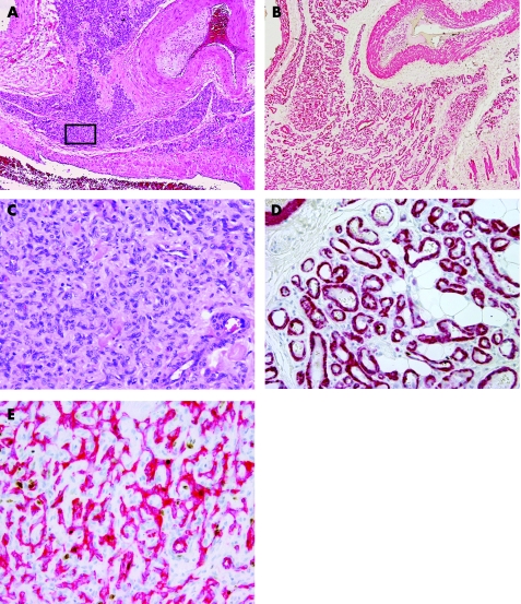
Figure 1 (A) Extensive (grade 3) solid microvascular proliferation amid the large vessels of an arteriovenous malformation (H&E). (B) Same area as in fig 3A showing distinct closely packed vascular structures in anti‐1a actin immunostain (SMA‐1). (C) Details taken from boxed area in fig 3A showing solid‐appearing growth of swollen vascular cells with inconspicuous lumina (H&E). (D) Details taken from boxed area showing well‐outlined vascular walls of small vessels in SMA‐1 immunostain. (E) Details taken from boxed area showing the luminal endothelial component of the vascular proliferation in anti‐CD31 immunostain.

An official website of the United States government
Here's how you know
Official websites use .gov
A
.gov website belongs to an official
government organization in the United States.
Secure .gov websites use HTTPS
A lock (
) or https:// means you've safely
connected to the .gov website. Share sensitive
information only on official, secure websites.
