Primary colonic glomus tumours are exceptionally rare and little is known about their natural history. Glomus tumours are mesenchymal tumours of pericytic origin, derived from the glomus body. They occur most commonly as benign subcutaneous lesions of peripheral soft tissue; however, they can infrequently arise at visceral sites including the stomach, pancreas, liver, lung, intestines and genitourinary tract.
We report the case of a previously well 37‐year‐old man who presented with a 3‐year history of intermittent abdominal pain and altered bowel habit. At colonoscopy a “cobblestoned” area in the ascending colon was encountered; biopsy showed a tumour of uncertain pathology but with no clear evidence of malignancy. A CT scan revealed no abnormality in this area and there was no radiological evidence of metastatic disease. After discussion, an extended right hemicolectomy was performed and the patient made an uncomplicated recovery.
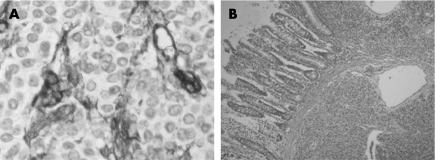
On gross examination, the tumour appeared flat and indurated; it was sited on the posterior wall of the ascending colon, measuring 30×20 mm. Microscopic examination of the tumour showed densely cellular lobules of varying size. The cells were small and round or cuboidal and intimately related to small vascular spaces within the lobules (fig 1A). No significant mitotic activity or necrosis was seen. The tumour involved the submucosa (fig 1B), muscularis propria and extended transmurally into the paracolic fat with prominent vascular invasion. Local lymph nodes in the resected specimen showed no evidence of tumour involvement.
Figure 1 (A) Immunostaining for CD34 shows regular cuboidal tumour cells surrounding prominent vascular channels. (B) Glomus tumour shown below the colonic mucosa.
Stains for mucin and neuroendocrine granules were negative. Immunohistochemistry was positive for vimentin and smooth muscle actin (SMA), but was negative for Cam5.2, CK7, CK20, LCA, chromogranin, synaptophysin, CD56, S100, desmin, CD117 and CD34. The histological and immunohistochemical profile confirmed the diagnosis of a primary colonic glomus tumour.
The immunohistochemical pattern and histological description are common to the three previously reported cases of colonic glomus tumours where data were presented,1,2,3 and share similar immunohistochemical profiles to both extra‐colonic gastrointestinal (GI) and peripheral glomus tumours.1 However, in previous reports, the colonic glomus tumours were intramural1 or confined to the adventitia2 or submucosa.3 Our case is unique in describing a tumour showing transmural growth extending into paracolic fat.
Regardless of the site of lesion in the GI tract, glomus tumours have many features which are usually associated with malignancy such as vascular invasion and focal atypia.1 However, this apparent malignant potential is not supported by the known natural history of colonic glomus tumours as the previously reported cases have always exhibited benign behaviour and metastatic disease has never been reported.1,2,3 By contrast, extra‐colonic GI glomus tumours have been seen to metastasise,1,4 and the best criteria to predict their malignant potential are those of Folpe et al, which are based on size (>2 cm), atypical mitotic activity, nuclear atypia and deep location.5 Miettinen et al found that GI and peripheral glomus tumours are histologically and immunohistochemically comparable, but recommended that prognostic comparison between these two groups was inadvisable.1
As so few colonic glomus tumours have been reported, therefore, the metastatic potential of these tumours remains uncertain but is probably very low.
Footnotes
Competing interests: None.
Informed consent was obtained for publication of the patient's details in this report
References
- 1.Miettinen M, Paal E, Lasota J.et al Gastrointestinal glomus tumours: a clinicopathologic, immunohistochemical, and molecular genetic study of 32 cases. Am J Surg Pathol 200226301–311. [DOI] [PubMed] [Google Scholar]
- 2.Barua R. Glomus tumour of the colon: first reported case. Dis Colon Rectum 198831138–140. [DOI] [PubMed] [Google Scholar]
- 3.Tuluc M, Abraham H, Inniss S.et al Case report: Glomus tumour of the colon. Ann Clin Lab Sci 20053597–99. [PubMed] [Google Scholar]
- 4.Kim J K, Won J H, Cho Y K.et al Glomus tumour of the stomach: CT findings. Abdom Imaging 200126303–305. [DOI] [PubMed] [Google Scholar]
- 5.Folpe A, Fanburg‐Smith J, Miettinen M.et al Atypical and malignant glomus tumours: analysis of 52 cases with proposal for the reclassification of glomus tumours. Am J Surg Pathol 2001251–12. [DOI] [PubMed] [Google Scholar]


