
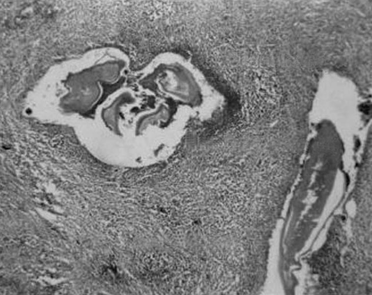
Figure 1 A photomicrograph showing dead hyalinised larvae along with their cuticles. The worms are surrounded by mixed inflammatory cell infiltrate, consisting mainly of lymphocytes, with few plasma cells and eosinophils (H&E staining, ×4).
Official websites use .gov
A
.gov website belongs to an official
government organization in the United States.
Secure .gov websites use HTTPS
A lock (
) or https:// means you've safely
connected to the .gov website. Share sensitive
information only on official, secure websites.
Figure 1 A photomicrograph showing dead hyalinised larvae along with their cuticles. The worms are surrounded by mixed inflammatory cell infiltrate, consisting mainly of lymphocytes, with few plasma cells and eosinophils (H&E staining, ×4).