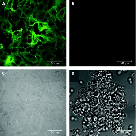
Figure 1 Immunofluorescence localisation of tissue factor (TF) expressed by HKC‐5 cells using confocal microscopy. (A) Anti‐TF monoclonal antibody visualised by fluorescein isothiocyanate conjugated secondary antibody. (B) Control, no primary antibody. (C) Differential interference contrast (DIC) image of cells shown in (B). (D) Specificity control using (virtually non‐TF expressing) leucocyte preparation: DIC overlaid on fluorescence.

An official website of the United States government
Here's how you know
Official websites use .gov
A
.gov website belongs to an official
government organization in the United States.
Secure .gov websites use HTTPS
A lock (
) or https:// means you've safely
connected to the .gov website. Share sensitive
information only on official, secure websites.
