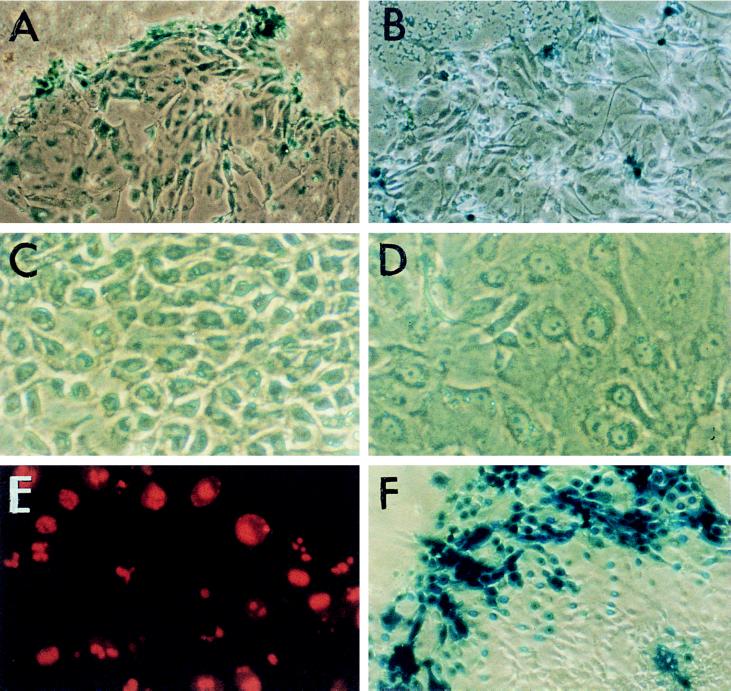
Figure 1.
Gene transfer into primary cultures of human airway epithelial cells. (A and B) Peripheral location of the transfected cells shown by the distribution of the X-Gal-positive cells after transfer of the E. coli lacZ gene by lipid BGTC (A) or by Transfectam (B). (×200.) (C and D) Phase-contrast micrographs showing the morphology of cultured airway epithelial cells located in the center (C) or at the periphery (D) of cell clusters. (×400.) (E) Localization of proliferating cells evidenced by immunofluorescent detection of Ki67 staining. (×400.) (F) Distribution of X-Gal-positive cells when AdRSVβGal adenoviruses were used at a high multiplicity of infection. (×200.)

