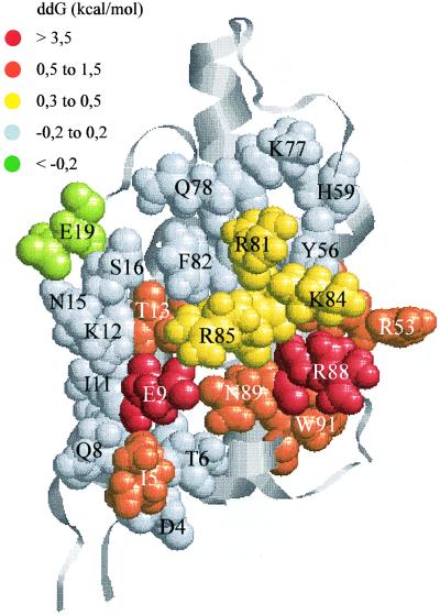
Figure 2.
Functional epitope of IL-4 determining the high-affinity binding of the receptor α chain. The 4-helix-bundle structure of IL-4 (17) is depicted as ribbons. The mutated residues are shown by space-filling models with their colors indicating the loss in binding free energy {ddG = 1.36 log(Kd[variant]/Kd[IL-4])} due to alanine or glutamine (E9, R53) substitution (see Table 2).

