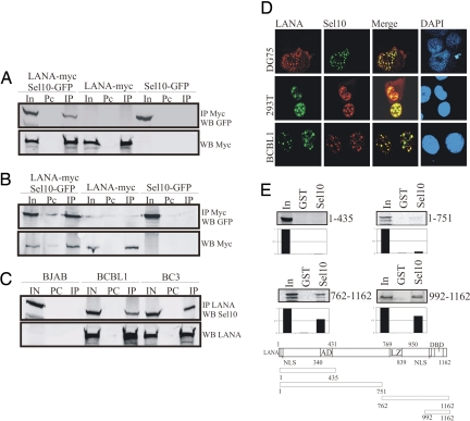
Fig. 1.
LANA associates with Sel10. (A and B) Immunoprecipitation analysis with mouse anti-Myc antibody or rabbit anti-GFP antibody showed that Sel10-GFP was directly immunoprecipitated with LANA-myc in 293T (A) and DG75 (B) cells. Fifteen million cells were cotransfected with 10 μg of Sel10-GFP and 10 μg of LANA-myc expression vectors or transfected with these vectors, respectively, 24 h after transfection; cell lysates were use for immunoprecipitate (IP) analysis. In, 5% total cell lysate input; Pc, preclear. (C) Endogenous LANA interacts with Sel10 in KSHV-infected primary effusion lymphoma cells BCBL1 and BC3. Immunoprecipitation analysis with polyclonal rabbit LANA antiserum showed that Sel10 was directly immunoprecipitated with LANA in BCBL1 and BC3 cells. For this experiment, 30 million cells were harvested for preparation of cell lysates, which were use for IP analysis. BJAB cells, which are KSHV-negative were used as a control. IN, 5% total lysate input; PC, preclear; IP, immunoprecipitate. (D) Immunofluorescence analysis showed that Sel10 was localized to the same nuclear compartment as LANA in different cells. (Top and Middle) DG75 and 293T cells were cotransfected with 10 μg each of Sel10-myc (Myc tagged) and pCDNA-LANA expression vectors, respectively; 24 h after transfection, cells were harvested for immunofluorescence analysis. Rabbit anti-LANA polyclonal antibody and mouse anti-Myc monoclonal antibody and corresponding secondary antibodies were used for this assay. (Bottom) BCBL1 cells were also used for immunofluorescence analysis. Human anti-LANA serum and rabbit anti-Sel10 polyclonal antibody and corresponding secondary antibodies were used for this assay. (E Top and Middle) C terminus LANA associates with Sel10 in vitro. The 35S-labeled products were incubated with GST as well as GST-Sel10 fusion protein. The pulldown products were electrophoresed on 10% SDS/PAGE gel, dried, and exposed to a PhosphorImager. Input controls of 10% of total LANA translation products. (Bottom) A schematic for the LANA clones used is given. NLS, nuclear localization signal; AD, acidic domain; LZ, leucine zipper; DBD, DNA-binding domain.

