Abstract
Detecting plant health conditions plays a key role in farm pest management and crop protection. In this study, measurement of hyperspectral leaf reflectance in rice crop (Oryzasativa L.) was conducted on groups of healthy and infected leaves by the fungus Bipolaris oryzae (Helminthosporium oryzae Breda. de Hann) through the wavelength range from 350 to 2 500 nm. The percentage of leaf surface lesions was estimated and defined as the disease severity. Statistical methods like multiple stepwise regression, principal component analysis and partial least-square regression were utilized to calculate and estimate the disease severity of rice brown spot at the leaf level. Our results revealed that multiple stepwise linear regressions could efficiently estimate disease severity with three wavebands in seven steps. The root mean square errors (RMSEs) for training (n=210) and testing (n=53) dataset were 6.5% and 5.8%, respectively. Principal component analysis showed that the first principal component could explain approximately 80% of the variance of the original hyperspectral reflectance. The regression model with the first two principal components predicted a disease severity with RMSEs of 16.3% and 13.9% for the training and testing dataset, respectively. Partial least-square regression with seven extracted factors could most effectively predict disease severity compared with other statistical methods with RMSEs of 4.1% and 2.0% for the training and testing dataset, respectively. Our research demonstrates that it is feasible to estimate the disease severity of rice brown spot using hyperspectral reflectance data at the leaf level.
Keywords: Hyperspectral reflectance, Rice brown spot, Partial least-square (PLS) regression, Stepwise regression, Principal component regression (PCR)
INTRODUCTION
Detecting plant health conditions plays a key role in farm pest management and crop protection (Everitt et al., 1994). Pathogens can cause severe damage to rice crops that often lead to reduced crop yield and quality. The use of large amounts of fungicide can effectively control most crop diseases and decrease the crop production loss. However, pesticide increases costs to growers while environmental problems may be incurred to soil, water, air and ecological systems (West et al., 2003). A proper on-time characterization and assessment of disease distribution and intensity could provide useful information for decision making about the timing of fungicide application in precise pest management.
In tradition, disease is detected visually by experienced growers who have the ability to detect subtle changes in plant color or a slight droop (wilt) or curl of plant leaves. Scout and plant canopy were recorded to assess the percentage of infection surface area in field (Jackson, 1986). However, this method is labor intensive and time consuming, and it is impossible to accurately estimate the infected areas and severity in large-scale farming (Kobayashi et al., 2001). Therefore, it is necessary to develop new effective and inexpensive approaches that can improve and supplement traditional techniques.
The developing remote sensing techniques make it possible to detect the changes in plants caused by disease stress (Brenchley, 1968). The crop disease assessment by remote sensing started about eighty years ago. In the late 1920’s, aerial photography was used to survey infection by Phymatotrichum omnivorum (Shear) Duggar in Texas (Neblette, 1927). Since then, a new era of disease detection by remote sensing started. In the subsequent several decades, aerial photography and satellite images were widely used to detect and characterize plant pests (Colwell, 1956; Jackson and Wallen, 1975; Nilsson, 1995; Mirik et al., 2006; 2007). However, most of these studies involved the use of airborne cameras and radiometers to record the reflected electromagnetic energy on analogue films that cover broad spectral bands (Huang and Apan, 2006).
Hyperspectral remote sensing is a new technique that utilizes sensors operating in hundreds of contiguous narrow wavelength bands which may have potential to improve the assessment of crop disease (Carter, 1994; Adams et al., 1999; Singh et al., 2007). Numerous studies demonstrate that hyperspectral reflectance and its detection accuracy of plant biochemicals were affected by the autocorrelation and multicollinearity of the data due to the continuous wavebands (Card et al., 1988; Yoder and Pettigrew-Crosby, 1995). It is unavoidable in the study of plant disease. Many research studies report that simple or multiple regressions do not show strong functional relationships between hyperspectra and plant health condition. Therefore, more sophisticated statistical approaches such as principal component regression (PCR), artificial neural networks (ANN), fuzzy systems, and partial least-square (PLS) regression analysis have been employed (Warner and Shank, 1997; Kimes et al., 1998; Muhammed and Larsolle, 2003). Nevertheless, the performance comparisons among regression (i.e. multiple stepwise regressions), PCR or PLS regression analyses were seldom conducted for the estimation of plant disease severity.
Rice brown spot is an aggressive plant disease caused by Bipolaris oryzae Shoem (Helminthosporium oryzae). It occurs in rice production areas all over the world and is one of the most common diseases in China. Rice brown spot causes severe damage under the conditions of cool summer and nitrogen deficiency, particularly in southern China. High humidity (>92.5%), leaf wetness and temperature (24 to 30 °C) are favorable conditions for disease development (Picco and Rodolfi, 2002). Wind and rainfall can spread the spores to other organs of the same individual and other plants. Losses can be severe if weather and field conditions are favorable for disease spreading. Rice brown spot can be found during the whole growing season. At the early stage, symptoms of brown spot mainly appear on the leaves. Leaf lesions reduce nutrient absorption and photosynthetic area, which result in the decrease of tillering nodes. There are no reports related to the use of hyperspectral reflectance data for the detection and measurement of rice brown spot disease.
The specific objectives of this research were threefold. The first objective was to confirm the possibility of detecting and measuring the rice brown spot disease at the leaf level by hyperspectral remote sensing. The second objective was to investigate the performance of multiple stepwise regression and PCR analysis or PLS regression for monitoring plant disease. Finally, to determine a reliable and stable statistical technique to estimate the disease severity of rice brown spot by reducing the autocorrelation and multicollinearity of hyperspectral reflectance.
MATERIALS AND METHODS
Study site and experiment design
The experiment was conducted at the village of Sankengkou, Wuyi County, Zhejiang Province, China. The study site is located at 28°42.013′ N latitude and 119°43.932′ E longitude, and at an altitude of 970 m above the sea level. Average annual rainfall is 1 477 mm. The growing season is generally short and lasts for 228 d. The mean annual temperature is 16.9 °C and the mean temperatures in January and July are 4.7 °C and 28.8 °C, respectively.
Seeds of rice were sown on June 5, 2006. Planting density was 25 seedlings/m2. Basal application of fertilizer of N, P2O5 and K2O was performed at 40, 30 and 30 kg/ha, respectively, with an additional application at 30, 20 and15 kg/ha respectively at the flowering stage. Rice brown spot disease occurred naturally in the field. Disease severity was determined by estimating the percentage of infected surface area of rice leaves in the laboratory.
Measurements of individual leaf reflectance
The fresh foliage from the field was placed in a special holder for measurements in the laboratory. Spectral measurements from individual leaves were made using the ASD Field Spectroradiometer Pro (Analytical Spectral Devices Inc., Colorado, USA). The wavelength of the measurement was configured for the range of 350 to 2 500 nm. The instrument has a spectral sampling resolution of 1.4 nm and a spectral interval of 3 nm between 350 to 1 000 nm, and a spectral sampling resolution of 2 nm and a spectral interval of 10 nm between 1 000 to 2 500 nm, respectively. Each leaf was positioned on a dark background. The field of the spectrometer view was 25° with 1 cm-diameter in the centre portion of the leaf. An incidence angle of 35° maintained at a standard distance (50 cm) was used throughout the study. The light source used was one 50 W halogen lamps. Upper leaf surface reflectance was observed. Each reading was the result of 10 replicate spectra that were averaged. All spectral measurements were made relative to a BaSO4 standard reference-panel, which was observed immediately before and after the spectral measurements of rice leaves.
Data processing and statistical methods
Reflectance data were firstly smoothed with a five point moving average to suppress instrumental and environmental noise before the data were further analyzed (Kobayashi et al., 2001). The total dataset consisted of 263 leaves which were divided into two subsets, one with 80% of the data was used as a training dataset for calibration (n=210) and the other with 20% used as testing dataset for validation (n=53). In this paper, the statistical methods were conducted with procedures in SAS 9.0 (Statistical Analysis System 9.0). These spectra represented the domains of 400~1 300, 1 501~1 780, and 2 051~2 350 nm, and the missing segments of 350~399, 1 301~1 500, 1 780~2 050, and 2 351~2 500 nm correspond to strong noise or water vapor absorption in the atmosphere and thus are not of interest for remote sensing of the Earth’s surface (Price, 1994).
In the agricultural sciences, the workhorse for statistical analysis has been multiple stepwise linear regression, although rationing of derivatives at various standard wavelengths has also been extensively used (Williams et al., 1983). Stepwise regression is a procedure for automatically selecting, in a stepwise manner based on partial correlations of a dependent variable with the independent variables that are close to optimal in the sense of maximizing the squared multiple correlations coefficient (R 2) of the dependent variable with the set of selected independent variables (Card et al., 1988). In the present context, the dependent variable is disease severity index of rice brown spot, and the independent variables are reflectance observed at a set of equally spaced wavelengths. The final regression equation was computed in Procedure STEPWISE in SAS 9.0. Several statistical papers have expressed concerns over the potentially large Type-I error by selecting independent variables that are merely noise, and inflating the R 2 of the stepwise procedure. The multiple stepwise linear regression does not have a strong theoretical basis and it cannot overcome the over-fitting and co-linearity of independent variables due to the high autocorrelation (Card et al., 1988). In our work, stepwise regression works quite well as long as the number of sample spectra is sufficiently large (n>200).
PCR is an effective data reduction technique that transforms the original dataset into a substantially smaller and easier to interpret set of uncorrelated variables. It has the capability of preserving the total variance while minimizing the mean square approximate errors, and is also used as a means to identify dominant modes of data that represent most of the information in the original dataset. Principal component analysis is quite useful for analysis of remote sensing data in which certain channels exhibit high degrees of dependence (Fung and LeDrew, 1987). Uncorrelated and linearly transformed components were computed from the original data in such a way that the first principal component accounts for the maximum proportion of the variance of the original dataset. Subsequent components were also calculated for the maximum proportion of the remaining variance in Procedure PCR in SAS 9.0. The PCR method has been applied to various remote sensing projects of plant disease involving in both field and laboratory spectral analysis (Holden and LeDrew, 1998). The original spectra were standardized by centering and scaling. An orthogonal transformation was applied to the correlation matrix in the study with component patterns that are visually interpretable. Although PCR selects factors that explain variation and predict plant biophysical or biochemical parameters by regression, it alleviates the high inter-band correlations of hyperspectral reflectance. However, the PCR method is not so convenient to selecting independent variables (spectral wavebands) without regard to the dependent variable (Wang, 1999).
The main goal of PLS procedure is to minimize the sample prediction error, seeking linear functions of the predictors that explain as much variation in each response as possible. In addition, it has the additional goal of accounting for variation in the predictors, under the assumption that directions in the predictor space which are well sampled should provide better prediction for new observations when the predictors are highly correlated. All of the techniques implemented in the PLS procedure, which are derived from PCR, are to reduce rank regression and PLS regression. The procedure works by extracting successive linear combinations of the predictors called either components or latent vectors to explain predictor variation. PLS regression is mainly used for modeling linear regression between multi-dependent variables and multi-independent variables. PLS not only avoids the harmful effects in modeling due to the multicollinearity when the inter-wavebands have strong autocorrelations, but also avoids over-fitting when the number of observations is less than the number of variables.
There are many reports about the application of PLS in agricultural science (Hansen et al., 2002), but few reports about its use for the detection and measurement of plant disease. One purpose of this study was to determine the efficiency of PLS procedure for detecting and estimating plant disease severity.
RESULTS
Multiple stepwise regression analysis for disease severity of rice brown spot
Multiple stepwise linear regressions were performed on the training dataset for disease severity of rice brown spot. The optimal number of variables or steps used by the stepwise regression procedure was seven, which yielded the best prediction for disease severity of rice brown spot. All wavelengths reflectance left in the model was significant at the 0.01 level. Wavelengths reflectance left in the final model was selected from the visible part of the spectra, which were located at 533, 567 and 693 nm.
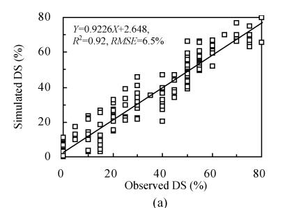
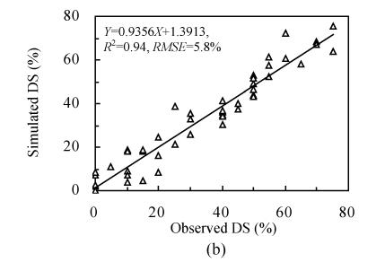
The coefficients of determination (R 2) between observed and predicted disease severity for the training and testing datasets were approximately 0.92 and 0.94, respectively (Table 1). The root mean square errors (RMSEs) of the training and testing dataset were 6.5% and 5.8%, respectively. Scatte plots between the observed and predicted disease severity on the training and testing datasets illustrate these results in Figs.1a and 1b.
Table 1.
Results of stepwise regression modeling disease severity of rice brown spot at the leaf level (P<0.01)
| RMSE (%) | R2 | n | |
| Training dataset | 6.5 | 0.923 | 210 |
| Testing dataset | 5.8 | 0.936 | 53 |
Fig. 1.
Observed vs predicted disease severity (DS) of rice brown spot using stepwise regressions (pace=7). (a) and (b) were obtained from the training and testing dataset, respectively
PCR for disease severity of rice brown spot
The results of the PCR revealed that the first eigenvector (PC-1, i.e. the first principal component) explained 78.34% of the variance of the original spectra data and the second component (PC-2, i.e. the second principal component) explained 12.04%. Although the third component could explain 4.9% of the variance of the spectra, it was considered to be low compared with the first two principal components. Therefore, the first two eigenvectors can be regarded as the principal components and the rest can be dropped from further analysis.
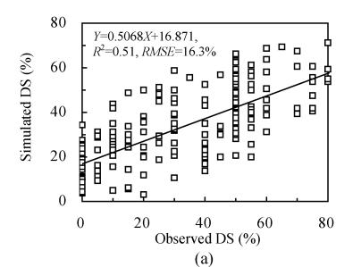
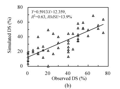
In this study, the first two principal components were used as the independent variables to build the regression model for estimating the disease severity of rice brown spot. The scatter plots of predicted vs observed disease severity for the training and testing dataset were shown in Figs.2a and 2b, respectively. The coefficients of determination (R 2) computed between observed and predicted disease severity for training and testing datasets were approximately 0.51 and 0.63, respectively. Meanwhile, low RMSEs of 16.3% and 13.9% were obtained for the training and testing dataset, respectively.
Fig. 2.
Observed vs predicted disease severity (DS) of rice brown spot using the first two principal components. (a) and (b) were obtained from the training and testing dataset, respectively
PLS regression for disease severity of rice brown spot
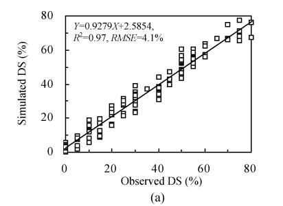
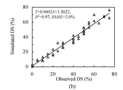
PLS regression was computed using the training dataset (n=210). Seven PLS factors were used to explain 98.56% of the model variance and 92.93% of the variation for prediction (Table 2). The seven-factor PLS model based on the training dataset was conducted for predicting disease severity of the testing dataset. The correlation coefficients between predicted and observed values for the calibration and validation sample were 0.986 and 0.985, respectively. The root mean square error of calibration (RMSEC) and validation (RMSEV) were 4.1% and 2.0%, respectively (Figs.3a and 3b). Clearly, the disease severity of rice brown spot was more accurate according to the RMSE values compared with other two methods.
Table 2.
Percent of variation accounted by partial least-square factors (n=210)
| Number of factors | Model effects (%) |
Prediction (%) |
||
| Current | Total | Current | Total | |
| 1 | 77.95 | 77.95 | 46.43 | 46.43 |
| 2 | 11.04 | 88.99 | 19.02 | 65.45 |
| 3 | 4.49 | 93.48 | 17.40 | 82.85 |
| 4 | 3.19 | 96.66 | 6.74 | 89.60 |
| 5 | 0.95 | 97.61 | 1.51 | 91.10 |
| 6 | 0.75 | 98.37 | 0.79 | 91.89 |
| 7 | 0.19 | 98.56 | 1.04 | 92.93 |
Fig. 3.
Observed vs predicted disease severity (DS) of rice brown spot using partial least-square regression. (a) and (b) were obtained from the training and testing dataset, respectively
DISCUSSION
In our study, three statistical methods consisting of multiple stepwise regression and principal component analysis or PLS regression analysis were investigated to ascertain their performances in the estimation of rice brown spot disease severity using remotely sensed leaf reflectance data.
Although the multiple stepwise linear regression technique has never had a strong theoretical basis (Card et al., 1988), it explained a large variation in the disease severity of rice brown spot. The three wavelengths in the visible range (533, 567 and 693 nm) were successfully selected to build the prediction model. These variables were neither related to the known water absorption wavebands at 970, 1 200, 1 400, 1 450, and 1 940 nm nor with the leaf pigment absorption wavebands at 430, 460, 640 and 660 nm (Curran, 1989). It was observed that wavelengths of 533 and 567 nm were located in the visible green region that is poorly absorbed by chlorophyll and other pigments, while 693 nm was located before the red-edge region where vegetation spectra are sensitive to variation of plant biochemistry (Vogelmann et al., 1993).
PCR was used successfully to determine the spectral properties of unhealthy plants with healthy ones (Holden and LeDrew, 1998). However, the low coefficients of determinant (R 2) and high prediction errors were obtained for both training and testing datasets, and the results suggested that the disease severity of rice brown spot was possibly influenced by other factors not explained by the first two principal components (Fig.2). The low prediction accuracy was similar to that obtained by Monteiro et al.(2007). These authors found that it was difficult to obtain a high level of accuracy with PCR method because it accepted independent variables (spectral reflectance) without considering the dependent variable (disease severity).
The high covariance and redundancy in the hyperspectral reflectance data influenced the prediction accuracy as reported by Thenkabail et al.(2000), and PLS models are ideal method for eliminating the problem. In our study, the high coefficients of determination and low prediction errors obtained with PLS regression for the training and testing dataset were corroborated by Hansen et al.(2002) and also by Huang and Apan (2006) for the prediction of grain yield and protein content using repeated canopy reflectance.
In conclusion, the performance of PLS regression analysis to estimate rice brown spot was better than multiple stepwise regression and PCR. Therefore, PLS can be regarded as the best suitable statistics approach for estimating disease severity of rice brown spot. The prediction errors estimated by the RMSEs ranged from 2.0% to 16.3% using the above statistical methods. We have demonstrated that it was feasible to estimate the disease severity of rice brown spot using hyperspectral reflectance data at the leaf level. It should be recognized that external factors such as wind, cloud and plant architecture bring much noise in the spectral reflectance at the canopy level. So these results obtained at the leaf level should be followed by field studies to take these factors into consideration.
Footnotes
Project supported by the Hi-Tech Research and Development Program (863) of China (No. 2006AA10Z203), and the National Science and Technology Task Force Project (No. 2006BAD10A01), China
References
- 1.Adams ML, Philpot WD, Novell WA. Yellowness index: an application of spectral second derivatives to estimate chlorosis of leaves in stressed vegetation. International Journal of Remote Sensing. 1999;20(18):3663–3675. doi: 10.1080/014311699211264. [DOI] [Google Scholar]
- 2.Brenchley GH. Aerial photography for the study of plant diseases. Annual Review of Phytopathology. 1968;6(1):1–22. doi: 10.1146/annurev.py.06.090168.000245. [DOI] [Google Scholar]
- 3.Card DH, Peterson DL, Matson PA. Prediction of leaf chemistry by the use of visible and near infrared reflectance spectroscopy. Remote Sensing of Environment. 1988;26(2):123–147. doi: 10.1016/0034-4257(88)90092-2. [DOI] [Google Scholar]
- 4.Carter GA. Ratios of leaf reflectance in narrow wavebands as indicators of plant stress. International Journal of Remote Sensing. 1994;15(3):697–703. doi: 10.1080/01431169408954109. [DOI] [Google Scholar]
- 5.Colwell RN. Determining the prevalence of certain cereal diseases by means of aerial photography. Hilgardia. 1956;26:223–286. [Google Scholar]
- 6.Curran PJ. Remote sensing of foliar chemistry. Remote Sensing of Environment. 1989;30(3):271–278. doi: 10.1016/0034-4257(89)90069-2. [DOI] [Google Scholar]
- 7.Everitt JH, Escobar DE, Summary KR, Davis MR. Using airborne video, global positioning system, and geographical information system technologies for detecting and mapping citrus blackfly infestations. Southwestern Entomologist. 1994;19(2):129–138. [Google Scholar]
- 8.Fung T, LeDrew E. Application of principal components analysis to change detection. Photogrammetric Engineering and Remote Sensing. 1987;53(12):1649–1658. [Google Scholar]
- 9.Hansen PM, Jørgensen JR, Thomsen A. Predicting grain yield and protein content in winter wheat and spring barley using repeated canopy reflectance measurements and partial least squares regression. Journal of Agricultural Science. 2002;139(3):307–318. doi: 10.1017/S0021859602002320. [DOI] [Google Scholar]
- 10.Holden H, LeDrew E. Spectral discrimination of healthy and non-healthy corals base on cluster analysis, principal components analysis and derivative spectroscopy. Remote Sensing of Environment. 1998;65(2):217–224. doi: 10.1016/S0034-4257(98)00029-7. [DOI] [Google Scholar]
- 11.Huang JF, Apan A. Detection of sclerotinia rot disease on celery using hyperspectral data and partial least squares regression. Journal of Spatial Science. 2006;52(2):129–142. [Google Scholar]
- 12.Jackson RD. Remote sensing of biotic and abiotic plant stress. Annual Review of Phytopathology. 1986;24(1):265–287. doi: 10.1146/annurev.py.24.090186.001405. [DOI] [Google Scholar]
- 13.Jackson HR, Wallen VR. Microdensitometer measurements of sequential aerial photographs of field beans infected with bacterial blight. Phytopathology. 1975;65(9):961–968. [Google Scholar]
- 14.Kimes DS, Nelson RF, Manry MT, Fung AK. Attributes of neural networks for extracting continuous vegetation parameters from optical and radar measurements. International Journal of Remote Sensing. 1998;19(14):2639–2663. doi: 10.1080/014311698214433. [DOI] [Google Scholar]
- 15.Kobayashi T, Kanda E, Kitada K, Ishiguro K, Torigoe Y. Detection of rice panicle blast with multispectral radiometer and the potential of using airborne multispectral scanners. Phytopathology. 2001;91(3):316–323. doi: 10.1094/PHYTO.2001.91.3.316. [DOI] [PubMed] [Google Scholar]
- 16.Mirik M, Michels GJJr, Kassymzhanova-Mirik S, Elliott NC, Catana V, Jones DB, Bowling R. Using digital image analysis and spectral reflectance data to quantify damage by greenbug (Hemitera: Aphididae) in winter wheat. Computers and Electronics in Agriculture. 2006;51(1-2):86–98. doi: 10.1016/j.compag.2005.11.004. [DOI] [Google Scholar]
- 17.Mirik M, Michels GJJr, Kassymzhanova-Mirik S, Elliott NC. Reflectance characteristics of Russian wheat aphid (Hemiptera: Aphididae) stress and abundance in winter wheat. Computers and Electronics in Agriculture. 2007;57(2):123–134. doi: 10.1016/j.compag.2007.03.002. [DOI] [Google Scholar]
- 18.Monteiro ST, Minekawa Y, Kosugi Y, Akazaw T, Oda K. Prediction of sweetness and amino acid content in soybean crops from hyperspectral imagery. ISPRS Journal of Photogrammetry and Remote Sensing. 2007;62(1):2–12. doi: 10.1016/j.isprsjprs.2006.12.002. [DOI] [Google Scholar]
- 19.Muhammed HH, Larsolle A. Feature vector based analysis of hyperspectral crop reflectance data for discrimination and quantification of fungal disease severity in wheat. Biosystems Engineering. 2003;86(2):125–134. doi: 10.1016/S1537-5110(03)00090-4. [DOI] [Google Scholar]
- 20.Neblette CB. Aerial photography for the study of plant diseases. Photo-Era Magazine. 1927;58:346. [Google Scholar]
- 21.Nilsson HE. Remote sensing and image analysis in plant pathology. Annual Review of Phytopathology. 1995;33(1):489–527. doi: 10.1146/annurev.py.33.090195.002421. [DOI] [PubMed] [Google Scholar]
- 22.Picco AM, Rodolfi M. Pyricularia grisea and Bipolaris oryzae: a preliminary study on the occurrence of airborne spores in a rice field. Aerobiologia. 2002;18(2):163–167. doi: 10.1023/A:1020654319130. [DOI] [Google Scholar]
- 23.Price JC. How unique is spectral signatures? Remote Sensing of Environment. 1994;49(3):181–186. doi: 10.1016/0034-4257(94)90013-2. [DOI] [Google Scholar]
- 24.Singh D, Sao R, Singh KP. A remote sensing assessment of pest infestation on sorghum. Advances in Space Research. 2007;39(1):155–163. doi: 10.1016/j.asr.2006.02.025. [DOI] [Google Scholar]
- 25.Thenkabail PS, Smith RB, Pauw ED. Hyperspectral vegetation indices and their relationships with agricultural crop characteristics. Remote Sensing of Environment. 2000;71(2):158–182. doi: 10.1016/S0034-4257(99)00067-X. [DOI] [Google Scholar]
- 26.Vogelmann JE, Rock BN, Moss DM. Red edge spectral measurements from sugar maple leaves. International Journal of Remote Sensing. 1993;14(8):1563–1575. doi: 10.1080/01431169308953986. [DOI] [Google Scholar]
- 27.Wang HW. Partial Least-Squares Regression—Method and Applications. Beijing: National Defense Industrial Press; 1999. pp. 150–170. (in Chinese) [Google Scholar]
- 28.Warner TA, Shank M. An evaluation of the potential for fuzzy classification of multispectral data using artificial neural networks. ISPRS Journal of Photogrammetric Engineering and Remote Sensing. 1997;63(11):1285–1294. [Google Scholar]
- 29.West JS, Bravo C, Oberit R, Lemaire D, Moshou D, McCartney HA. The potential of optical canopy measurement for targeted control of field crop diseases. Annual Review of Phytopathology. 2003;41(1):593–614. doi: 10.1146/annurev.phyto.41.121702.103726. [DOI] [PubMed] [Google Scholar]
- 30.Williams PC, Norris KH, Gehrke CW, Bernstein K. Comparison of near-infrared methods for measuring protein and moisture in wheat. Cereal Foods World. 1983;28(2):149–152. [Google Scholar]
- 31.Yoder BJ, Pettigrew-Crosby RE. Predicting nitrogen and chlorophyll content and concentrations from reflectance spectra (400~2500 nm) at leaf and canopy scales. Remote Sensing of Environment. 1995;53(3):199–211. doi: 10.1016/0034-4257(95)00135-N. [DOI] [Google Scholar]


