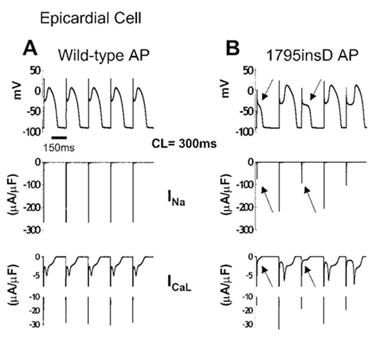
Figure 5.

AP morphology changes in 1795insD epicardial cells. The 96th through 100th beats (CL = 300 ms) are shown for a simulated epicardial cell. A, WT APs (top) with INa (middle) and ICaL (bottom). B, 1795insD APs (top) with corresponding INa and ICaL. Beat-dependent reduction in mutant INa (middle, arrows) on the background of large Ito and IKs present in epicardial cells acts to deepen the AP notch, which suppresses plateau ICaL activation (bottom, arrows), resulting in loss of the AP dome (top, arrows).
