
Figure 4.
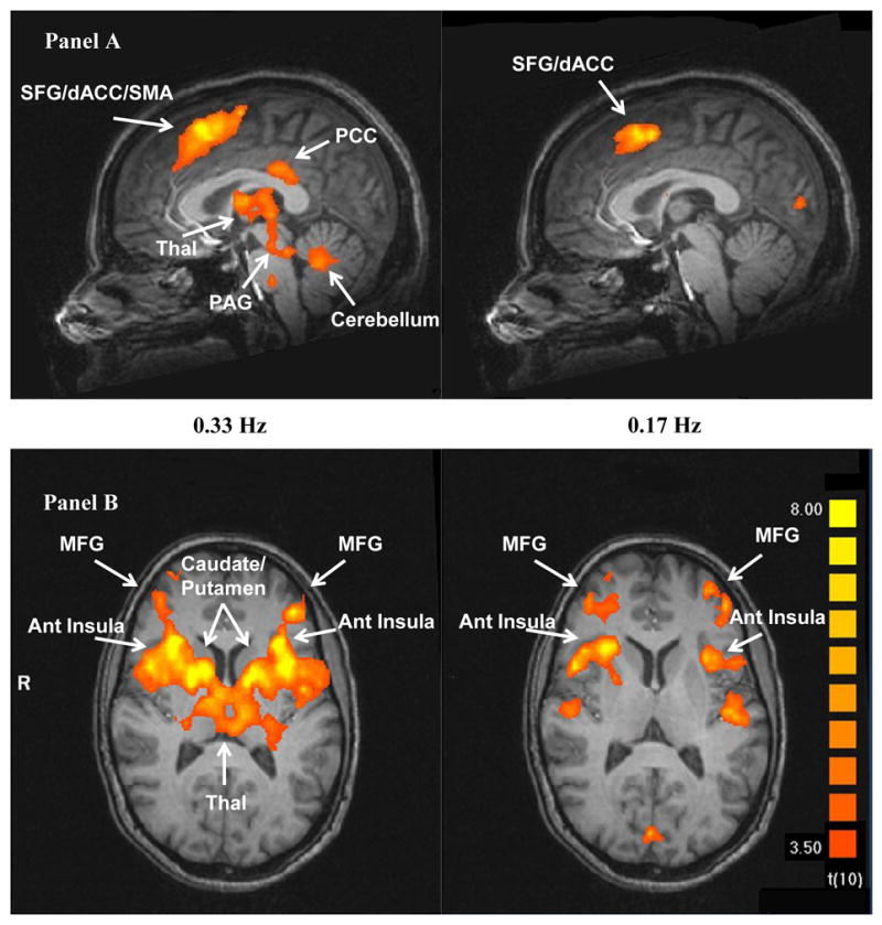
Pain related brain activity of the last of six heat pulses at 0.33 Hz and 0.17 Hz in sagittal (Panel A) and transverse (Panel B) sections, respectively. In contrast to Fig. 3 which illustrates the relationship between brain activity and the number of heat pulses, theses fMRI images depict the association of brain activity with TSSP pain ratings. Significantly increased brain activation was detected in PFC, SMA, ACC, THAL, ACC, and INS (p < .005).