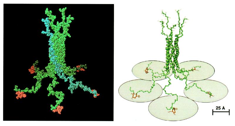
Figure 6.
Space-filling (Left) and ribbon (Right) representations of a model of the three-dimensional structure of Pab-S. Binding peptides are in red. The upper part of the structure shows six histidine residues at each C terminus. One chain within the pentameric molecule is highlighted in blue in the space-filling representation. Five shaded circles (radius of 40 Å) under the ribbon structure schematically denote receptor molecules. The ribbon representation was generated with the program molscript (28).

