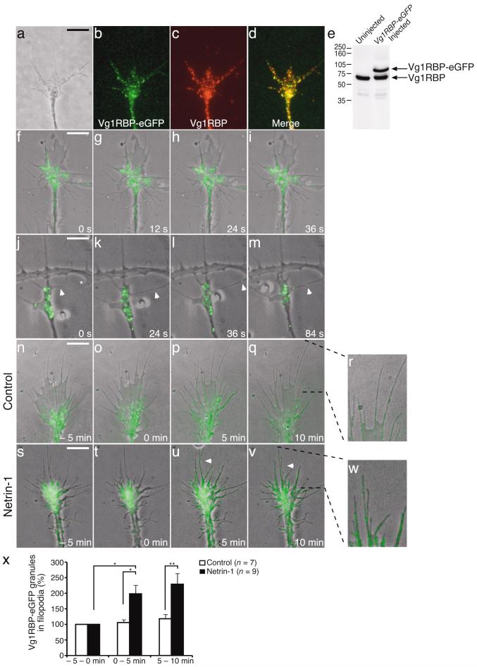
Figure 2. Netrin-1 or cell-contact induce translocation of Vg1RBP-eGFP into filopodia.
Retinal explants taken from stage 33/34 embryos expressingVg1RBP-eGFP were stained with Vg1RBP antibody. Both endogenous and overexpressed proteins are recognized by the Vg1RBP antibody and show similar patterns of localization (a: phase, b: Vg1RBP-eGFP, c: Vg1RBP, d: merge). Vg1RBP-eGFP is expressed at similar levels as the endogenous Vg1RBP as shown by Western blot analysis of head lysates from injected and uninjected embryos (e). Time-lapse analysis of Vg1RBP-eGFP shows bidirectional movement of Vg1RBP-eGFP granules in the axon shaft, growth cone central domain and filopodia. Images were taken every 12 seconds (f-i and Supplementary movie 1). Vg1RBP-eGFP granules are also detected moving along filopodial contact-contact sites (j-m, arrowheads and Supplementary movie 2). The asterisk indicates the branch/filopodium, which is going to contact the Vg1RBP-eGFP expressing filopodium. Live imaging of Vg1RBP-eGFP expressing retinal growth cones stimulated with control medium (n-r) or netrin-1 (s-w and Supplementary movie 3) at T = 0 min. The percentage of Vg1RBP-eGFP granules in the filopodia was calculated in each frame and averaged per 5 minutes (x). Application of netrin-1 results in an increase of Vg1RBP-eGFP granules in the filopodia compared to control. * P < 0.05, ** P < 0.01, Kruskal-Wallis test. Error bars s.e.m. Scale bars 5 μm.

