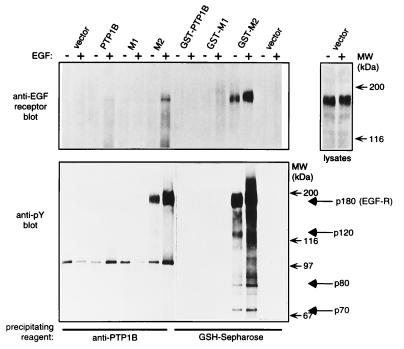
Figure 3.
Identification of tyrosine-phosphorylated p180 in the D181A precipitates as the EGFR. This experiment was conducted as described in the legend of Fig. 2 except that the resulting immunoprecipitates were split in half, subjected to electrophoresis on duplicate SDS/polyacrylamide gels, and blotted with either anti-EGFR mAb (Transduction Laboratories) (Upper Left) or with anti-pTyr (4G10) (Lower Left). Pairs of lanes represent samples from duplicate transfections. A − indicates that for 24 h prior to harvesting the cells were maintained in media without serum. A + indicates that these cells were similarly deprived of serum until 10 min before harvesting, during which time they were incubated with EGF at 100 ng/ml. The anti-pTyr blot was deliberately overexposed in an attempt to observe pTyr-containing proteins in the C215A (M1) precipitates and to visualize p70 and p80 in the D181A (M2) precipitates. Anti-EGFR staining was weaker than anti-pTyr staining of p180 in D181A (M2) precipitates, presumably either because the anti-EGFR antibody is of lower affinity or because there are multiple pTyr epitopes recognized by 4G10 in the “trapped” EGFR. (Right) Anti-EGFR blot of 50 μg of lysate from untransfected COS cells that were untreated (−) or incubated for 10 min with EGF at 100 ng/ml (+).

