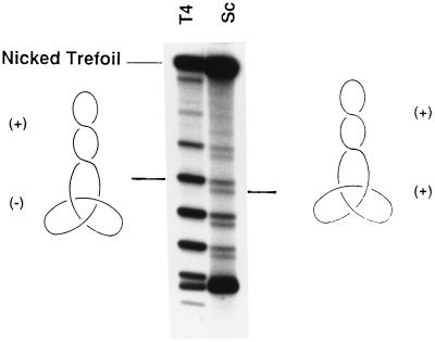
Figure 3.
A single ladder of positively supercoiled topoisomers prepared from the negative-noded trefoil formed by treatment of negatively supercoiled pCC56cos with phage T4 DNA topoisomerase (left lane, T4). For comparison, the sample analyzed in lane 3 of Fig. 2, which was prepared by converting nicked trefoil formed by S. cerevisiae DNA topoisomerase II to the positively supercoiled form, was run again in the right lane (Sc). The drawings to the left and right schematically depict the sign of the knot nodes and that of crossings in the plectonemically wound supercoiled DNA. The single ladder of topoisomers in the left lane comigrate with the trailing ladder of topoisomers in the right lane.

