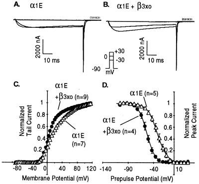
Figure 2.
Effect of β3xo (xo28) on α1E. (A and B) Time courses of activation of α1E in oocytes injected with α1E cRNA alone or cRNAs encoding both α1E and β3xo (clone xo28). (C and D) Effect of β3xo on voltage-dependent activation and inactivation of α1E. G–V curves were obtained from peak tail currents measured by stepping to −50 mV after depolarizing pulses of 25-msec duration from −88 to 116 mV in 4-mV increments. The data were sampled at 10 kHz and filtered at 2 kHz. The data points were fitted by the sum of two Boltzmann distributions. For α1E, the first component had a V1/2 = 2.7 mV, an effective valence (zδ) = 2.9 e, and a relative amplitude of 51%; the second had a V1/2 = 41.8 mV and a zδ = 1.4 e. For α1E + β3xo, the first component had a V1/2 = 2.7 mV, a zδ = 3.4 e and a relative amplitude of 75%; the second had a V1/2 = 51.8 mV and a zδ = 1.3 e. Steady-state inactivation curves were derived from peak currents elicited by a pulse to +20 mV following a conditioning pulse of 10 sec to potentials from −120 to 27 mV in 7 mV increments and a brief (4-msec) pulse to −90 mV. The data were sampled at 500 Hz and filtered at 100 Hz. Sweeps were separated by 20 sec to allow a full recovery from inactivation. Data points were fitted by a Boltzmann distribution. The effective valences were 2.7 ± 0.1 e for α1E and 3.4 ± 0.1 e for α1E + β3xo; the half-inactivation potentials were −32.2 ± 3.4 mV (n = 5) for α1E and −53.5 ± 1.1 mV (n = 4) for α1E + β3xo.

