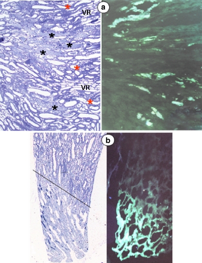
Fig. 7.
Characteristic morphological findings in rats subjected to the ARF protocol and injected with DDC. Cryo-sections from the left kidney were used for DDC fluorescence analysis, whereas the in vivo fixed right kidney was used for light microscopy morphological examination of methylene blue-stained slides. Analyses of both kidneys were performed in a blinded fashion. An example is shown of the striped pattern reflecting the damage gradient, where tubules adjacent to the vasa recta (VR) are preserved (red stars) while those distant from vasa recta are injured (black stars) (a, left panel, ×100). Note the excellent co-localization of morphologically damaged tubules with the DDC fluorescence (a, right panel, ×100) in the contralateral kidney. DDC fluorescence was selectively identified in all morphologically injured regions, including papillary tip structures (b: left panel ×40, right panel ×100)

