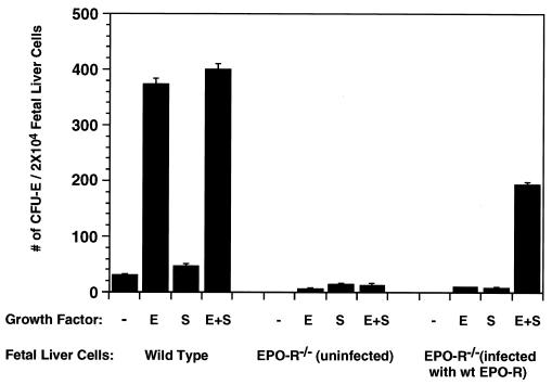
Figure 1.
Formation of erythroid colonies by wt and Epo-R−/− fetal liver cells. (Left) CFU-E colonies formed by wt fetal liver cells. (Center) CFU-E colonies formed by uninfected Epo-R−/− fetal liver cells. (Right) CFU-E colonies formed by Epo-R−/− fetal liver cells following infection with a retrovirus expressing the wt Epo-R cDNA. The average numbers of CFU-E colonies from three independent assays were expressed per 2 × 10⁴ nucleated fetal liver cells.

