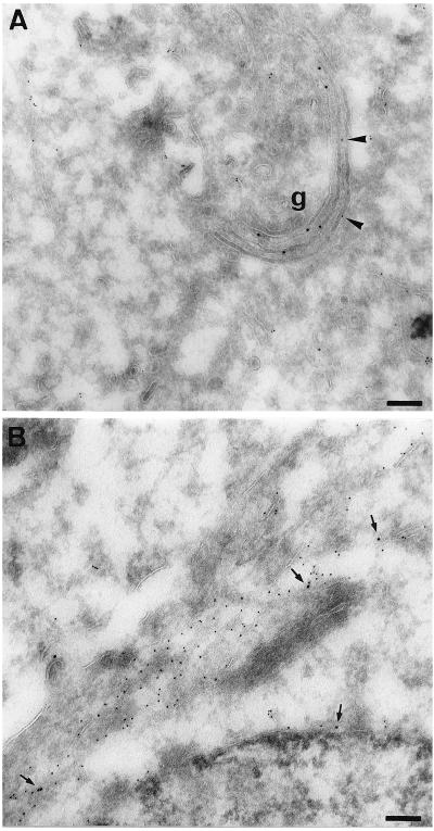
Figure 2.
Colocalization of gal-T and Ii in rab6 Q72L transfected cells. HeLa cells were cotransfected with Ii and either pGEM-1 (A) or rab6 Q72L (B) plasmids. Cells were fixed 6 h after transfection and processed for cryosectioning and immuno-electron microscopy. Cells were double labeled with antibody against gal-T and anti-Ii antibody. Primary antibodies were visualized with protein A-gold conjugates (gal-T, 10 nm; Ii, 5 nm). In control cells (A), anti-gal-T antibody labels typical Golgi stacks (g). Arrowheads in A point to Ii molecules localized in Golgi stacks in control cells. Arrows in B point to gal-T molecules localized within ER cisternae (Ii-positive) in rab6 Q72L transfected cells. (Bars = 0.1 μm.)

