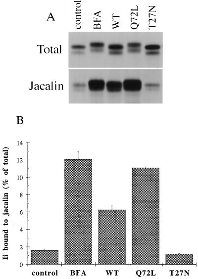
Figure 4.
rab6 Q72L, wt rab6, and BFA increase O-glycosylation of Ii. HeLa cells cotransfected with Ii and either empty plasmid (control, BFA) or the indicated rab6 constructs (WT, Q72L, T27N) were metabolically labeled for 10 min and chased for 3 h. In BFA-treated cells, 5 μg/ml BFA was added to the chase medium. Ii immune precipitates were solubilized with SDS and nine-tenths of each eluate was diluted in Triton X-100 containing buffer and incubated with biotinylated jacalin and streptavidin-agarose. O-glycosylated Ii bound to jacalin was specifically eluted with galactose. (A) Visualization by autoradiography of Ii present in the different cell lysates before (Total) or after binding to jacalin (Jacalin). Total represents one-tenth of the whole immunoprecipitate for each sample. (B) Ii bands were quantified by scanning with the PhosphorImager. This graph represents the means ± SD of two independent experiments.

