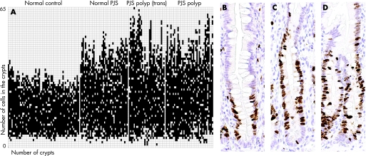
Figure 1 Ki67 staining pattern in intestinal crypts of normal and polyp tissue from patients with Peutz–Jeghers syndrome (PJS) and controls. In all samples the number and location of Ki67 positive cells was scored in 10 completely visible and longitudinally sectioned crypts. The proliferative compartment was defined as the cellular compartment extending between the cell positions of the lowermost and uppermost Ki67 positive labelled cell using the bottom of the crypt as the reference cell position 0.8 (A) Schematic overview of Ki67 staining, black = positive Ki67 staining. (B) Ki67 staining on normal control intestine. (C) Ki67 staining on PJS normal intestine. (D) Ki67 staining on PJS polyp.

An official website of the United States government
Here's how you know
Official websites use .gov
A
.gov website belongs to an official
government organization in the United States.
Secure .gov websites use HTTPS
A lock (
) or https:// means you've safely
connected to the .gov website. Share sensitive
information only on official, secure websites.
