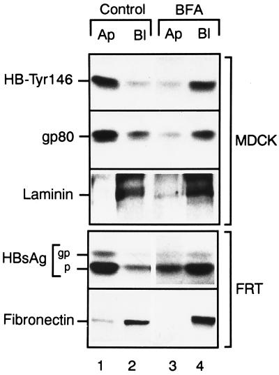
Figure 5.
HBsAg and HB-Tyr146 were secreted predominantly basolaterally when the apical pathway was blocked by BFA. MDCK and FRT cells grown in Transwell chambers were pulse-labeled with 35S-labeled methionine and cysteine for 30 min and chased for 6–8 h. Immunoprecipitations from apical and basolateral media of laminin, HB-Tyr146, and gp80 secreted by MDCK cells, and fibronectin and HBsAg secreted by FRT cells were made. Control conditions (lanes 1 and 2) showed a preferential apical secretion of HBsAg and the N-glycosylation negative mutant. Transfected MDCK and FRT cells still possess an intact basolateral pathway, disclosed by their secretion of laminin and fibronectin, respectively. BFA (10 μg/ml), added 30 min before pulse labeling and maintained during the chase (lanes 3 and 4), did not affect basolateral secretion of these proteins, whereas apical secretion of HBsAg and HB-Tyr146 was inhibited, redirecting them to basolateral.

