Figure 2.

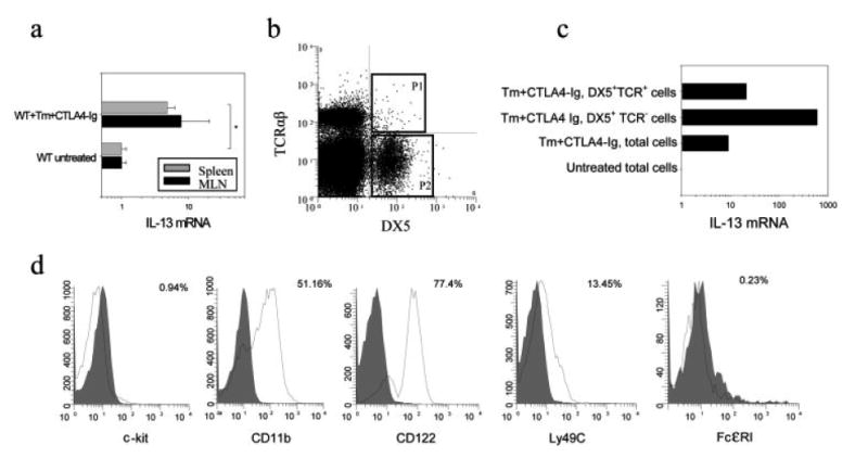
NK cells are a major non-T cell source of IL-13. BALB/c mice were inoculated orally with T. muris (Tm) eggs and treated with CTLA4-Ig as previously described. At day 21 after inoculation (a) MLN and spleens were assayed for IL-13 gene expression as described in Fig. 1, and (b) spleen cells from CTLA4-Ig-treated infected mice were depleted of CD4+ T cells by magnetic bead cell sorting and then stained with PE-labeled anti-mouse TCRαβ and FITC-labeled anti-mouse DX5 mAb. DX5+TCRαβ+ cells (gate P1) and DX5+TCRαβ− (gate P2) cells were purified by electronic cell sorting. (c) Isolated DX5+TCRαβ+ and DX5+TCRαβ− cells were analyzed for IL-13 gene expression. RNA from total spleen cells in untreated mice was used as a control. (d) DX5+TCRαβ− cells from spleens were analyzed using multi-color FACS staining. Single-color histograms represent the expression of c-kit, CD11b, IL-2Rβ, Ly49C and FcεRI on gated DX5+TCRαβ− cells (solid line). Corresponding isotype controls are shown in gray. Asterisk indicates a statistically significant difference (*p<0.05) between the indicated groups.
