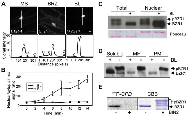
Figure 1. BR Induces Nuclear Localization of BZR1.
(A) Effect of brassinazole (BRZ) and brassinolide (BL) on the subcellular localization of BZR1-YFP. Transgenic Arabidopsis expressing BZR1-YFP were grown on MS or MS + 2 μM BRZ medium in the dark for 4 days. Seedlings grown on BRZ medium were treated with mock or 100 nM BL for 1 hr and images of the YFP signal (top) were obtained using confocal microscopy. Numbers in each image show the average ratios between nuclear and cytoplasmic signal intensities and standard errors calculated from seven cells for each treatment. The white lines inside the images show the areas used for line scan measurements that yielded plot profiles shown in the lower panels. N, nuclear signal, C, cytoplasmic signal. Fluorescent intensity is 1000x and scale bar is 10 μm.
(B) Kinetics of the BL-induced nuclear accumulation of BZR1-YFP. BZR1-YFP seedlings grown on 2 μM BRZ were treated with BL or mock solution (−BL), and images were acquired at each time point to measure the nuclear/cytoplasmic ratios. Error bars are ± standard errors of mean.
(C) Unphosphorylated BZR1 is enriched in the nuclear fraction. Immunoblot of unphosphorylated and phosphorylated BZR1-CFP proteins in total and nuclear fractions from mock- or BL-treated seedlings.
(D) Phosphorylated BZR1 is more enriched in membrane fractions than unphosphorylated BZR1. Transgenic plants expressing the BZR1-CFP protein were treated with (+BL) or without (−BL) 1 μM BL, and the soluble, microsomal (MF), and plasma membrane (PM) fractions were analyzed by immunoblot.
(E) BIN2 phosphorylation inhibits DNA binding activity of BZR1. Gel blot of unphosphorylated and BIN2-phosphorylated MBP-BZR1 proteins was probed with radiolabeled CPD promoter DNA. CBB, Coomassie Brilliant Blue stained gel. Unphosphorylated BZR1 (BZR1) and phosphorylated BZR1 (pBZR1) proteins are marked by arrows (C-E).

