
Figure 4-10.
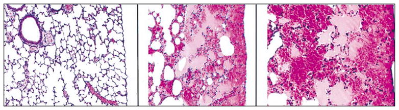
Left: Sham exposed control mouse lung (low magnification) exhibiting no lesions (H&E stain). Center: Mouse lung exposed to 30 kHz, CW ultrasound for 5 min (low magnification). Alveoli are contiguous with the visceral pleura and are filled with blood. The hemorrhage spreads to a finite limit into adjacent lung parenchyma (H&E stain). Right: CW exposed mouse lung (high magnification) exhibiting alveolar hemorrhage (H&E stain). The visceral pleura and contiguous alveolar septa are intact. (Photo courtesy of James F. Zachary, DVM, PhD.)