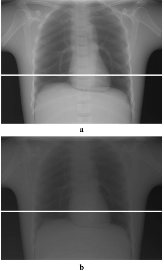
Fig. 3.

(a) Slot-scan and (b) open-field images of an anthropomorphic chest phantom shown in inverted log scale with same window and level settings. A bright line (in the image shadow of tungsten bar) across the lower lung and heart regions indicates where the scatter measurement took place.
