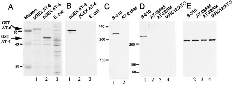
Figure 1.
Characterization of ATM protein antiserum pAb 132. (A) Lysates from E. coli transformed with the plasmids pGEX AT-6 (lane 1) and pGEX AT-4 (lane 2) following fusion protein induction and untransformed E. coli (lane 3) were subjected to SDS/PAGE and Coomassie blue staining. Migration of the specific GST–AT fusion proteins are indicated by arrowheads. (B) Extracts shown in A were immunoblotted with pAb 132. (C) A total of 50 μg of B-310 (lane 1, wild type for ATM) and AT24RM (lane 2, homozygous mutant for ATM) cell extracts were immunoblotted with pAb 132. (D) A total of 50 μg of SDS-lysate from B-310 lymphoblasts (lane 1) and A-T patient lines AT29RM (lane 2), AT22RM (lane 3), and IARC12/AT3 (lane 4) were immunoblotted with pAb 132. (E) Blot displayed in D was stripped and reprobed with an antisera to NuMA.

