Figure 5.
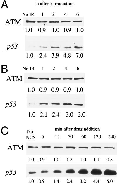
Effects of genome damaging agents on cellular ATM levels. (A) Hs-68 fibroblasts were exposed to 5 Gy of ionizing radiation. Unirradiated cells (No IR) and cells 1, 2, 4, and 6 h after exposure were analyzed for ATM protein (Upper) and p53 (Lower) levels by immunoblotting. (B) B-310 lymphoblasts were exposed to 5 Gy of IR and analyzed as in A. (C) The normal human fibroblast cell line F-2054 was treated with NCS and extracts from treated cells at 5, 15, 30, 60, 120, and 240 min following drug addition, and untreated cells (No NCS) were analyzed for ATM protein (Upper) and p53 (Lower). Relative immunoblot signal intensity (determined by densitometric scanning of exposed films) is displayed below each band. Minor differences in recorded ATM protein levels are likely due to slight inconsistencies in protein loading and/or protein transfer.

