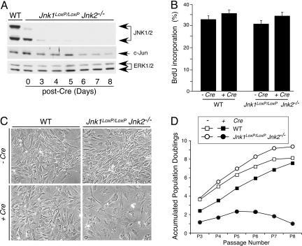
Fig. 2.
JNK deficiency causes a delayed reduction in cellular proliferation. (A) Time-course analysis of JNK expression after gene ablation. Jnk1LoxP/LoxP Jnk2−/− MEF were transduced by using a retroviral Cre expression vector. Cell extracts were prepared on different days after infection and examined by immunoblot analysis using antibodies to JNK, cJun, and ERK. Extracts prepared from WT MEF were also examined. (B) The effect of retroviral tranduction of Cre in WT and Jnk1LoxP/LoxP Jnk2−/− MEF was examined 10 days after infection by measurement of the incorporation of BrdU by flow cytometry. (C) The morphology of WT and Jnk1LoxP/LoxP Jnk2−/− MEF at passage 7 after retroviral transduction of Cre was examined by phase-contrast microscopy. (Magnification: ×100.) (D) The growth of WT and Jnk1LoxP/LoxP Jnk2−/− MEF was examined by using a 3T3 assay. The data are presented as the accumulated population doublings during eight passages in culture. The effect of retroviral transduction of Cre was investigated.

