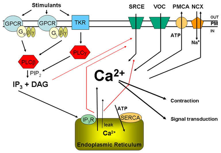
Figure 1.

Schematic diagram showing the major components that control [Ca2+]i in myometrium. These include the contractant hormone pathways that stimulate G-protein coupled (GPCR) and tyrosine kinase (TyKR) receptors, G-proteins composed of Gα subunits (Gαq, Gαi and (not shown) Gαh), the associated Gβ and Gγ subunits, phospholipase C β and γ, which convert PIP2 to IP3 and diacylglycerol (DAG). IP3 binds to IP3 receptors (IP3R) in the endoplasmic reticulum (ER), releasing Ca2+ from this intracellular store. Another ER release mechanism involves ryanodine receptors (RyR). Other Ca2+ entry mechanisms include cation channels responsive to stimuli such as IP3R activation, DAG and release of Ca2+ from the ER (SRCE) and voltage-operated Ca2+ channels (VOC). The plasma membrane Ca2+ ATPase (PMCA) and the Na/Ca exchanger (NCX) are responsible for moving Ca2+ out of the cell; the ER Ca2+ ATPase (SERCA) pumps Ca2+ back into the ER. There is also a passive leak of Ca2+ from the ER. Ca2+ has stimulatory effects on the contractile apparatus, resulting in contraction, and also acts as an intracellular signal influencing a number of pathways in the myometrium.
