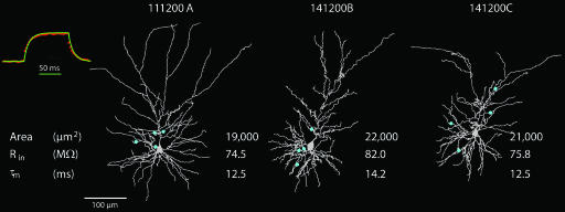
Fig. 1.
Three reconstructed pyramidal L2/3 pyramidal cells from the barrel cortex. The dendrites of the pyramidal cells are in gray and, in each case, the putative synaptic contacts established with specific presynaptic L4 spiny neurons are marked by blue dots. The input resistance (Rin) and membrane time constant (τm) are extracted from experimental transients measured in these cells. The membrane area of the dendritic tree (including spines) is also denoted. (Inset) Voltage trace after a 100-ms current step in the passive model of cell 111200A (green continuous line) superimposed on the averaged and normalized experiment voltage traces measured in this same cell (red dotted line).

