
Figure 8.
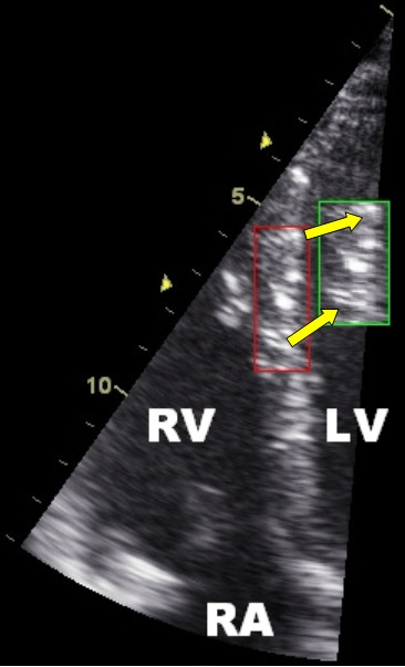
Typical speckled pattern of the myocardium on ultrasound in the septal wall. The tracking algorithm follows a unique speckle pattern during the cardiac cycle. The red square represents the starting location and the green square the location of the pattern at end systole. Note the change in distance between the speckles due to deformation (longitudinal shortening and radial thickening of the square). RV = right ventricle; LV = left ventricle; RA = right atrium.