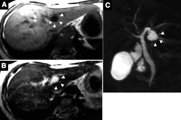
Figure 2.

Magnetic resonance imaging (MRI) reveals the cystic lesion as low in the T1-weighted image (A) and as high in the T2-weighted image (B). MR cholangiography shows a cystic lesion at the left lobe of the liver, but a filling defect in the bile duct and a communication between the cystic lesion and bile duct could not be defined (C). Arrows head indicate the cystic lesion.
