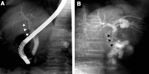
Figure 4.

Endoscopic retrograde cholangiography (A) shows a dilated common bile duct with defined filling defects corresponding to mucin. Percutaneous transhepatic cholangiography (B) also shows mucin in the common bile duct and a communication between the cystic lesion and bile duct. However, the filling defect corresponding to the tumor component in the cystic lesion could not be defined. Arrows head indicate mucin in the common bile duct.
