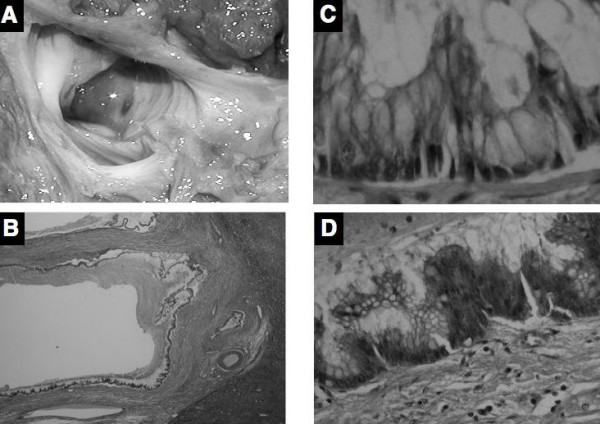
Figure 5.

(A) The gross appearance of the resected specimen does not show a mass protruding into lumen in the cystic lesion with mucin. (B) Microscopically, the cystically dilated bile duct does not have a mass of protruding lesion composed of papillary growth with fibrovascular cores or villous structures (hematoxylin and eosin ×40). (C) Cyst wall was lined by tall columnar epithelium with mucin hypersecretion (Hematoxylin and eosin ×200). (D) Neoplastic cells with hyperchromatic nuclei and loss of cell polarity was occasionally observed, but stromal invasion was not present (Hematoxylin and eosin ×200).
