


Figure 13.
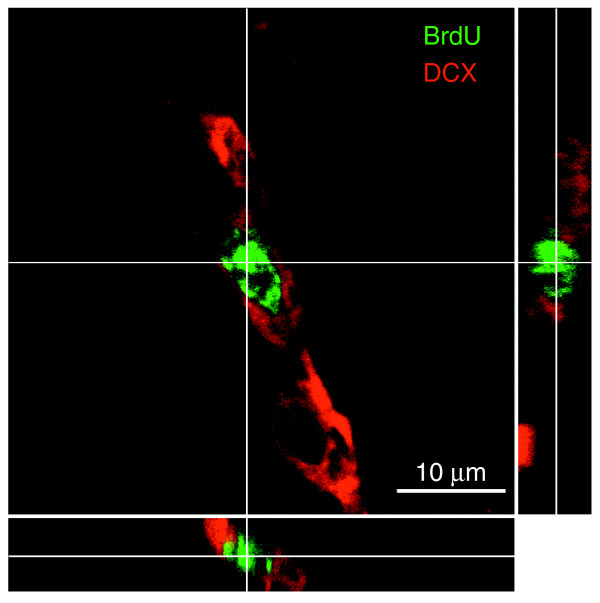
Slice (three-sided) reconstruction of a BrdU+/DCX+ cell. Photographs were taken at 2 μm intervals. Identical analyses were conducted for every cell that was scored as BrdU+ and expressing a cell-type specific antigen, as shown in Figure 4. The three-sided reconstruction shows that the BrdU+ nucleus (green) belongs to the DCX+ cell (red).