
Figure 6.
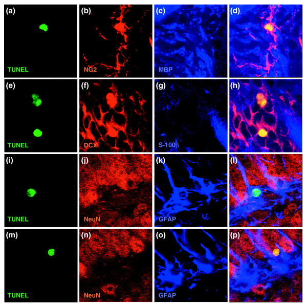
Representative images of co-labeling for TUNEL and expression of cell type-specific antigens. Despite the apparent labeling of nuclei with cell-type specific antibodies in dying cells (presumably due to the changes in antigen distribution associated with nuclear fragmentation), co-labeling was highly cell-type specific (see also Figure 7 for z-stack analysis). (a-d) NG2+/TUNEL+ cells from the CC. In this and subsequent rows, the first image is of TUNEL staining, the next two images are of staining for the proteins indicated, and the merged image is on the far right. (e-h) DCX+/TUNEL+ cells from SVZ; (i-l) GFAP+/TUNEL+ cell from DG. (m-p) NeuN+/TUNEL+ cell from DG. In all merged images except (l) co-labeled cells show up as yellow; in (l) the nucleus of the co-labeled cell is green. Magnification 400×.