
Figure 9.
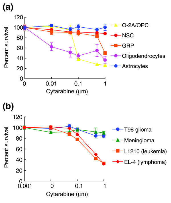
Primary CNS cells are equally or more vulnerable to cytarabine than cancer cells. Cells were plated on coverslips in 24-well plates at a density of 1,000 cells per well and allowed to grow for 24–48 h. On the basis of drug concentrations achieved in human patients, cells were exposed to cytarabine for 24 h. Cell survival and viability was determined after additional 24–48 h (see Materials and methods). (a) Rat neural cell types studied included O-2A/OPCs, oligodendrocytes, GRP cells, NSCs and astrocytes. (b) We also examined the T98 glioma cell line, a meningioma cell line, and the L1210 and EL-4 leukemia cell lines. To define the onset of cytarabine toxicity, cells were treated with cytarabine over a wide dose range (0.01–1 μM) extending downwards from the lower ranges achieved in high-dose therapy. Each experiment was carried out in quadruplicate and was repeated multiple times in independent experiments. Data represent mean of survival ± SEM, normalized to control values. There are no concentrations of cytarabine at which tumor cell lines were more sensitive O-2A/OPCs or oligodendrocytes.