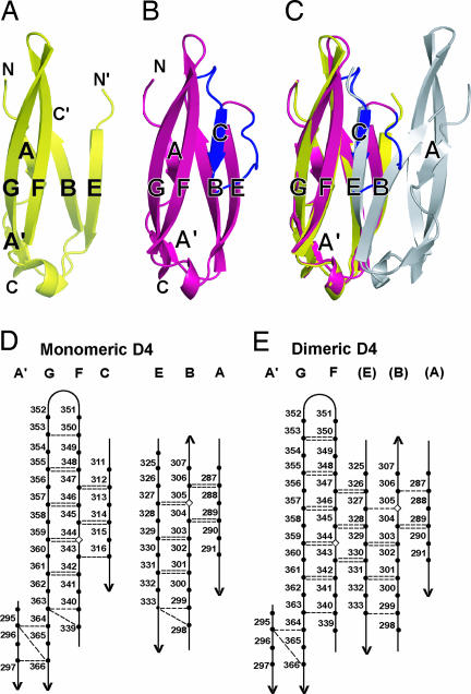
Fig. 2.
Structural differences between monomeric and dimeric D4. (A–C) Ribbon diagrams of D4 after superposition on D4 of one monomer of dimeric ICAM-1. (A) One monomer of dimeric D4. (B) Monomeric D4. (C) Superposition of monomeric and dimeric D4. The 16 residues that become ordered in monomeric D4 are shown in blue, and the residues preceding and following the disordered region in dimeric ICAM-1 are marked C′ and N′, respectively. (D and E) β-sheet hydrogen bonds in monomeric (D) and dimeric (E) D4. Backbone hydrogen bonds of ≥1.0 kcal/mol as determined with DSSP (29) are shown as dashed lines. The disulfide-bonded cysteines in β-strands B and F are shown as diamonds.

