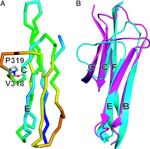
Fig. 3.
Structural properties of D4. (A) Backbone Cα trace of D4 colored in rainbow from highest (red) to lowest (blue) B factor. Atoms of cis-Pro-319 and atoms C and O of Val-318 are represented with sticks, and the hydrogen bond in the turn between the C-strand and CE meander is dashed. The disulfide bond is shown in yellow. (B) Comparison of the CE edges of D2 (magenta) and monomeric D4 (cyan) of ICAM-1. Superposition is on β-strands B, C, E, and F and the region containing β-strands A, A′, and G is omitted for clarity.

