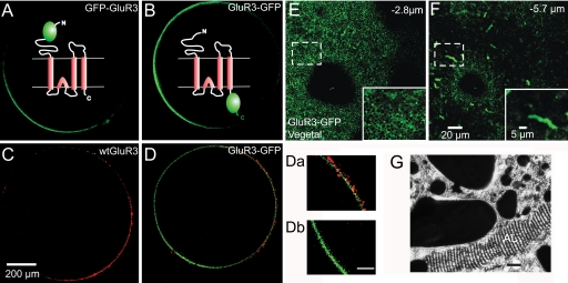
Fig. 1.
Visualization of GFP-tagged GluR3. (A–D) Confocal sections of oocytes injected in the equator with the indicated cRNAs, taken at 300 μm from the bottom of the oocyte. The animal hemisphere is at the right and the vegetal at the left. Note that the fluorescence is more intense in the vegetal hemisphere. Diagrams in A and B represent the GluR3 chimeras showing the extracellular or intracellular localization of the GFP. (C and D) Immunolocalization of GluR3 receptors in the plasma membrane of nonpermeabilized oocytes. (C) Oocyte expressing WT GluR3 incubated with an antibody against the amino terminus of GluR3, which is localized extracellularly and a secondary antibody (Alexa 568, red label). (D) Oocyte expressing GluR3-GFP incubated with the same primary and secondary antibodies. Note that the insertion of GluR3 is mainly polarized to the plasma membrane in the animal hemisphere. (Da and Db) Magnification of the membrane near the animal and vegetal poles of the oocyte shown in D. (Scale bar: 50 μm.) (E and F) Z-axis sequential scans in the vegetal hemisphere of an oocyte injected with GluR3-GFP. (Insets) Magnifications of the areas within the smaller squares. Images taken at 2.8 and 5.7 μm from the oocyte's surface. Below the surface, notice high fluorescence in elongated structures, resembling annulate lamellae. (G) Electron microscope image of an annulate lamellae (AL). (Scale bar: 1 μm.) Adapted from R.M. and C. Tate (unpublished work 1978).

