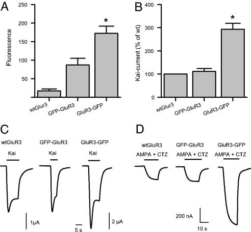
Fig. 3.
GluR3 GFP-constructs have different expressional potencies. (A) Fluorescence intensity (arbitrary units) of injected oocytes. The fluorescence of oocytes expressing WT GluR3 is the same as the native fluorescence observed in noninjected oocytes (n = 9 each). The fluorescence of oocytes expressing GluR3-GFP was double of that of oocytes expressing GFP-GluR3 (P < 0.05). (B) GluR3-GFP injected oocytes generated nearly three times more 100 μM Kai-current than oocytes injected with WT GluR3 or GFP-GluR3 cRNA (P < 0.05; n = 64). (C) Sample membrane currents evoked by 100 μM kainate in injected oocytes. The 2 μA calibration bar applies only to the GluR3-GFP current. (D) The currents elicited by 10 μM AMPA plus 10 μM CTZ were also larger in oocytes expressing the GluR3-GFP.

