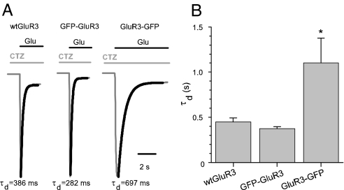
Fig. 6.
Carboxyl-terminal GFP tagging alters the decay of GluR3 receptors. (A) Glutamate (1 mM) plus CTZ (10 μM)-currents in oocytes injected with GluR3 or the GFP constructs. For better comparison, only the temporal course of activation and decay of the currents are shown. The inactivation was not complete after 20 s of agonist perfusion. After washing out the Glu plus CTZ, the currents returned to their basal level. (B) GluR3-GFP-currents decayed more slowly: τd = 1,102 ± 276 ms; n = 10; *, P < 0.05; vs. WT GluR3 (447 ± 44 ms; n = 10) and GFP-GluR3 (374 ± 20 ms; n = 12). Thicker traces are the exponential fits to the Glu-current traces. Currents were normalized for comparative purposes. Glu-current amplitudes were as follows: 600 nA for WT GluR3, 980 nA for GFP-GluR3, and 3,180 nA for GluR3-GFP.

