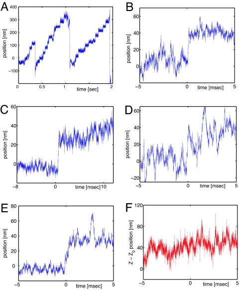
Fig. 2.
Myosin V stepping. (A) A single myosin V moving against the optical tweezers. In this run the measured stall force is 1.6 pN. (B–D) Examples of single 36-nm steps, extracted from the curve in A. (E and F) Bead motion during a single step. The trajectory is projected along the x axis (parallel to the actin filament) and the z axis (perpendicular to the actin filament).

