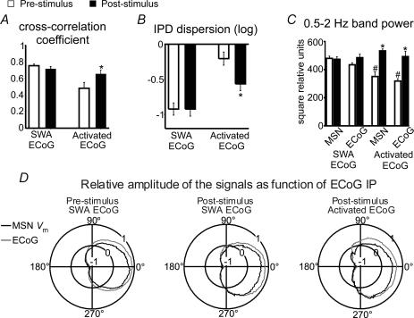
Figure 4. Effects of cortical stimulation on corticostriatal coupling.
The effect of cortical stimulation on the degree of coupling between the ECoG and MSN membrane potential was estimated by computing pre- and post-stimulus cross-correlograms and instantaneous phase differences (IPD). In the slow wave activity background condition (SWA ECoG), peak cross-correlation coefficients (A) and IPD circular dispersion (B) did not significantly differ between before and after stimulation (paired t test, n = 13). Conversely, in the activated ECoG condition (activated ECoG), peak cross-correlation coefficients increased (*P < 0.006) and IPD circular dispersion decreased (*P < 0.01) compared with pre-stimulus values (n = 8, paired t test), indicating that cortical stimulation enhanced low frequency coupling between cortical and striatal activity. For individual neurons, pre-stimulus cross-correlations in the SWA background condition were significant at P < 0.001 and non-significant for the activated ECoG background condition. C, cortical stimulation had no effect on ECoG or MSN membrane potential power when background activity was dominated by slow waves, but increased significantly low frequency signal components in both areas when background activity was activated (*P < 0.001 versus pre-stimulus condition, n = 8, paired t test). Both the ECoG and MSN Vm powers were significantly smaller in the activated ECoG condition compared with the SWA background (#P < 0.001, t test). D, polar plots depicting mean relative amplitudes (radial axis: −1 to +1) of MSN membrane potential (black) and ECoG (grey) as a function of ECoG instantaneous phase (IP) (angular axis: 0 to 360 deg). Signal amplitudes changed in parallel, with transitions from silent to active states taking place at ∼270 deg phase, both during spontaneous activity or after cortical stimulation and regardless of the background ECoG condition.

