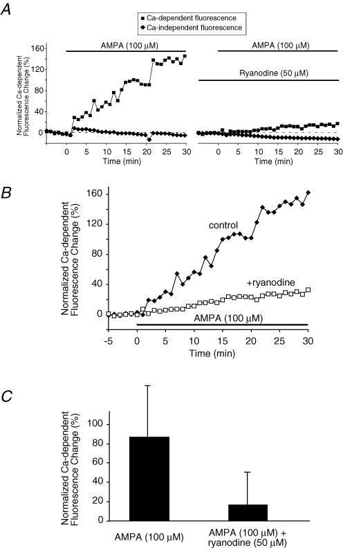
Figure 6. AMPA increases axoplasmic [Ca2+] in spinal cord dorsal column axons measured using confocal microscopy.
A and B, example time courses from one experiment of axoplasmic Ca2+ levels as reported by fluorescence change of Oregon Green-488 BAPTA-1 in normal, non-ischaemic dorsal column axons. AMPA (100 μm; plus 30 μm cyclothiazide to reduce desensitization) induced a substantial Ca2+ rise which was significantly blunted by ryanodine. A, shows Ca2+-dependent (Oregon Green-488 BAPTA-1) and Ca2+-independent (Alexa Fluor 594) fluorescence separately. B, illustrates the ratio of Ca2+-dependent to Ca2+-independent fluorescence to correct for the small downward drift in fluorescence over time. C, bar graph of mean axoplasmic Ca2+ fluorescence changes at 30 min. Fluorescence increased by a mean of 86 ± 53% above baseline after 30 min of AMPA exposure. Addition of ryanodine (50 μm) reduced the AMPA-evoked Ca2+ increase to 16 ± 34% (P < 10−7). These data suggest that AMPA receptors increase axonal [Ca2+] partly by controlling release of Ca2+ from ryanodine receptor-dependent axonal Ca2+ stores (see text). n = 48 and 26 axons for AMPA and AMPA + ryanodine, respectively.

