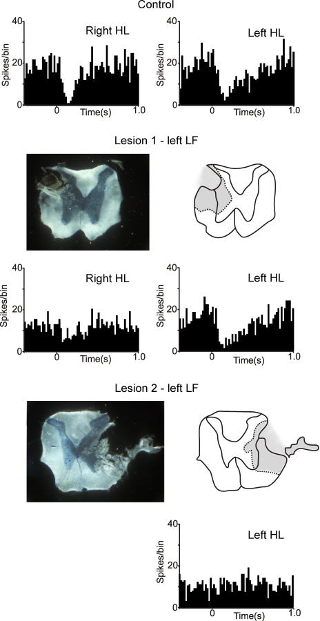
Figure 4. Effects of lateral funiculi lesions on Golgi cell long-lasting depressions.
The PSTHs show the responses of a Golgi cell with the cord intact (top row), after a lesion of the left lateral funiculi (LF; lesion 1, middle row) and after a more extensive lesion of the right LF (lesion 2; bottom row). Lesion 1 had little effect on the responses evoked by stimulation of the left hindlimb (HL; ipsilateral to the lesion) or the left FL, but reduced the responses to the right HL (by about 50%). Lesion 1 included the dorsal part of the LF, but spared some of the ventral part. After lesion 2, which removed all of the LF, the response evoked from the left HL (contralateral to lesion 2) was abolished. PSTHs built from 100 stimuli, bin-size 20 ms.

