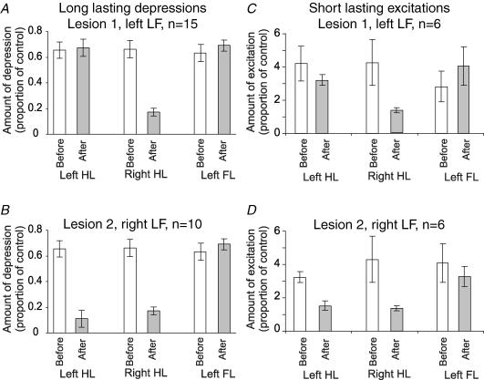
Figure 6. Effects of LF lesions on Golgi cell responses: grouped data.
A and C, mean responses of Golgi cells before (open bars) and after a lesion of the left LF (shaded bars). Responses to the left hindlimb and forelimb (left HL, left FL) were unaltered, but the responses to stimulation of the right hindlimb (right HL) were significantly attenuated (P < 0.001, paired t test). B and D, effects of a subsequent lesion of the right LF, after which not all of the Golgi cells remained discriminable. In this case the responses evoked by stimulation of the left HL were significantly reduced (P < 0.002, paired t test), while the responses to stimulation of forelimb afferents remained unaltered.

