
Fig. 3.
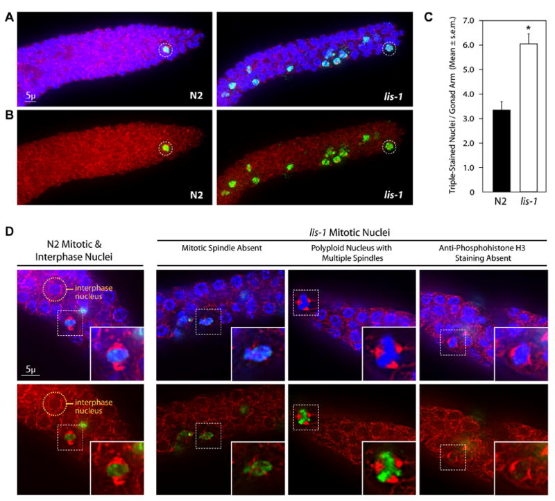
lis-1(lf) disrupts the mitotic spindle and produces cell cycle arrest. (A) lis-1(n3334) and N2 gonads stained with Hoechst, α-tubulin antibody, and α-phosphohistone H3 antibody (α-PH3). lis-1 gonads contain an increased number of α-PH3-positive nuclei compared to N2 gonads. Representative mitotic nuclei are circled for both strains. Photographs represent a z-stack through the width of the gonad, flattened using deconvolution microscopy. (B) Photographs of the same animals presented in (a), showing only α-tubulin antibody and α-PH3 staining. (C) The mean number of triple-stained nuclei per distal gonad arm is significantly greater in lis-1(n3334) than in the N2 control (P<0.0001, unpaired t-test). The data are averages ± s.e.m. of N2 (n=47) and lis-1 (n=61). (D) N2 mitotic nuclei were easily distinguished by their symmetrical bipolar spindles and distinct centralized Hoechst and α-PH3 staining. An N2 interphase nuclei is circled above for comparison. lis-1 nuclei, on the other hand, display a range of defects including spindle abnormalities, polyploidy, and lack of α-PH3 staining. Representative nuclei are indicated by dashed-squares and are magnified in the inset images.